Retina
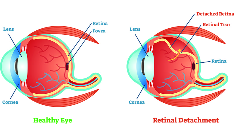
 The retina is a thin layer of tissue located along the back of the eye, near the optic nerve. Within the retina are light-sensitive cells called rods and cones — these cells receive light that enters the eye through the lens and translate it into neural signals. These signals are then sent to the brain via the optic nerve, allowing you to see.
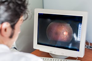
When damage occurs to the retina, it can cause individuals to lose their ability to see vivid colors or details, and may lead to vision loss in one or both eyes. It is important to schedule an eye exam as often as medically necessary, in order to identify and treat potential issues before they worsen. Some common diseases and conditions known to affect the health and function of the retina are listed below. At Prestera Eye, our skilled eye doctors can assess your specific concerns and create a customized treatment plan that can preserve the health of your retina and help you maintain clear vision.
- Diabetic Retinopathy
- Hypertensive Retinopathy
- Macular Degeneration
- Retinal Holes and Tears
- Retinal Detachment
- Retinal Vein Occlusion
Diabetic Retinopathy
Diabetic retinopathy is a disease that can affect individuals with diabetes when blood sugar levels are not properly maintained, damaging the blood vessels in the retina and ultimately causing impaired vision or blindness. Signs and symptoms of diabetic retinopathy are often difficult to detect early on, making routine eye exams crucial for those with diabetes.
Hypertensive Retinopathy
Similar to diabetic retinopathy, hypertensive retinopathy causes damage to the blood vessels in the retina, and can eventually cause blindness. Both conditions are progressive and have some of the same symptoms, but hypertensive retinopathy occurs when the blood vessels are narrowed, due to high blood pressure. This can limit proper retinal function and cause one’s vision to deteriorate over time. Scheduling regular eye exams is the best way to ensure this condition is identified and treated early on, before permanent damage can be done.
Macular Degeneration
 Macular degeneration is one of the leading causes of blindness in people over the age of 50. It is often referred to as Age Related Macular Degeneration, or AMD. The macula is a small spot located near the center of the retina, and it can begin to break down over time. When this happens, blurriness and blind spots may manifest, and vision becomes increasingly worse. Another common symptom of AMD is subretinal neovascularization, which is when new blood vessels are formed in the eye and cause hemorrhaging or leakage. Approximately 80% of the critical vision loss associated with macular degeneration can be attributed to subretinal neovascularization.
Retinal Holes and Tears
A retinal hole or tear can occur if the fluid inside the eye (known as the vitreous) begins to pull away from the retina, stretching it and causing it to tear in areas where it may be weak. In some cases, vision may not be affected right away, but it can cause symptoms such as blurriness, floaters, or flashes of light. Retinal tears typically are not painful, so without regular eye exams, these tears might not be identified until there has been serious damage — if fluid seeps through the hole and gets behind the retina, it can lead to retinal detachment, which requires immediate attention to avoid permanent vision loss.
Retinal Detachment
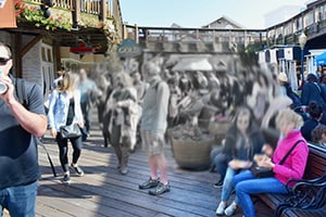
As noted above, a retinal detachment is a very serious condition that renders the retina unable to properly function. A retinal detachment should be considered an emergency, and needs to be treated as soon as possible. As the retina lifts away from the back of the eye, it can no longer process light and transmit neural signals through the optic nerve, ultimately causing visual distortions. If left untreated, a retinal detachment can cause permanent blindness. Signs of a retinal detachment include diminished peripheral vision, floaters, flashes of light, blurred vision, and a “curtain” or shadow effect within your field of vision. If you are experiencing these symptoms, please contact your eye doctor immediately.
Retinal Vein Occlusion
A retinal vein occlusion (RVO) is when a blood clot prevents a retinal vein from draining blood from the retina. This can cause hemorrhages and leakage of fluid, leading to vision loss. There are two types of RVO — central retinal vein occlusion (CRVO) and branch retinal vein occlusion (BRVO). CRVO is the blockage of the main retinal vein, whereas BRVO refers to when a smaller branch vein becomes blocked. If fluid begins to leak into the macula, it can swell, causing a macular edema, resulting in vision loss. These complications can often be treated with either injections of medicine into the eye or a retinal laser. RVO can also result in neovascularization and neovascular glaucoma — both of which lead to vision loss. If RVO is not properly treated, it can cause permanent blindness.
Contact Prestera Eye Medical Group
If you are experiencing any visual disturbances like the ones mentioned above, please contact us today to make an appointment with one of our skilled and knowledgeable eye doctors. Dr. Prestera or Dr. Guan can conduct a thorough eye exam and determine the best course of action to preserve your vision.